Patient Preparation
Procedure performed under anaesthetic
According to patients, the location, and the extent of angioma cavity. Surgery is performed under local or general anesthesia in an operating room equipped with a medical imaging device such as a fluoroscope. The medical imaging device will allow the practitioner to follow his procedure in live under fluoroscopic control.
Before any treatment, once the patient is under general sedation, the practitioner evaluates:
- The rate of blood flow in the cavity and prevents opportunities of angioma escape towards sensitive organs and tissues. These escapes are seen in live on the monitor of fluoroscope with a water-soluble contrast agent that is injected into the cavity.
- The size and depth of the angioma cavity to treat.

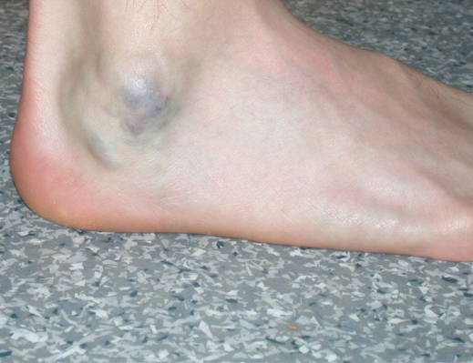
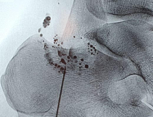
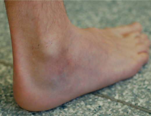
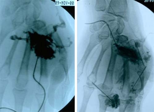
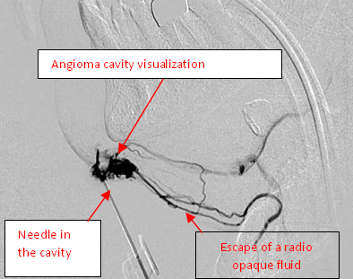
These escapes are visible live on the image intensifier monitor thanks to a water-soluble contrast agent that is injected into the cavity.
Once these safety conditions are met for the patient. The practitioner can proceed to treatment that is to say to the injection of SCLEROGEL kit®
Injection of SCLEROGEL kit
SCLEROGEL kit® injection under fluoroscopic control
Connect the 1 ml syringe containing the ethanol gel on the tubing of the winged infusion set inserted in the vein requiring treatment.
Unclamp the catheter tubing and push the ethanol gel relatively quickly so that it fills the tubing, to avoid blood possibly flowing into the tubing.
Once the ethanol gel has reached the end of the needle, inject it very slowly into the lumen requiring treatment.

Précision : On the fluoroscope monitor, thanks to the angiography, the low blood flow venous cavity appears of a homogeneous grey color (water-soluble agent). Progressively where the ethanol gel is going to progress in this venous cavity, the water-soluble contrast agent is going to be repulsed and to make place to the non radio opaque ethanol gel and this cavity will appear then white (colorless).
You are going to discover a certain number of results obtained with SCLEROGEL kit®
For the following pictures, in order to identify the presence of the gel in the angioma cavity, different agents of contrast have been mixed with the gel. It is not about a current use for the administration of SCLEROGEL kit®.

Before treatment

Water soluble contrast agent SCLEROGEL kit® mixed with a contrast agent to show its presence in the angiomatous cavity

After treatment

Before treatment

Water soluble contrast agent SCLEROGEL kit® mixed with a contrast agent to show its presence in the angiomatous cavity

After treatment
With the courtesy of Drs. DOMPMARTIN & LABBE – CHU CAEN – FRANCE Copyright©